
Page 94 - The Indian Optician Digital Edition January-February 2022
P. 94
CASE STUDY
ON THE EVALUATION OF
CORNEAL ENDOTHELIAL CELL
CHANGES IN PATIENTS
he cornea is a transparent, avascular, watch
glass-like structure. The normal value of corneal
Tendothelium is 2500 to 3500 cells/mm2. When
some corneal endothelial cells die and disappear due
to ageing, trauma or other stress such as contact
BRAHAMDEV MANDAL lens wear, the remaining cells are unable to divide
OPTOMETRIST fast enough to replace the dead cells. Instead, they
(B.OPTOM, M.OPTOM) enlarge and spread to cover the dead cells in order
LAHAN EYE CARE AND RESEARCH to maintain the intact monolayer mosaic; the neural
CENTRE, DHARBHAGA, BIHAR
crest uniformly distributes over the cornea.
Loss or damage of endothelial cells leads to an
increase in corneal thickness, which may ultimately
induce corneal decompensation and loss of vision.
The corneal endothelial cells play an important role
in regulating stromal hydration and maintaining the
transparency of the cornea by constantly removing
the fluid out of the corneal stroma. This function is
executed by active metabolic pumps in the corneal
endothelium. Normal corneal hydration represents
a balance between the leak across the endothelium
and the movement of water by the metabolic pump.
The status of the Corneal Endothelium Morphology is
usually described by the below aspects:
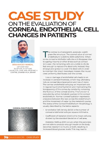
CORNEAL ENDOTHELIUM
− Endothelial Cell Density (ECD) is the number of
corneal endothelial cells per square millimetre.
− Coefficient of Variation (CoV) is the mean cell area
divided by the standard deviation of cell area.
Diabetes Mellitus (DM) occurs when the pancreas
are not able to produce enough insulin or the body
becomes resistant to insulin, or both, resulting in
increased blood glucose levels. This may lead to
| JAN-FEB 2022 | 90 CASE STUDY